Magnetic Resonance (MRI) is a diagnostic technique based on the use of magnetic fields which do not therefore use ionizing radiation. v2
With this technique we can see the hydrogen atoms of our body, which is what we are principally made up of (water, fat, proteins, etc.). So it is a technique that allows us to evaluate all the tissue of our body and detect disorders.
This technique is used a lot in the study of the Central Nervous System (brain and spinal cord) and when studying the locomotor apparatus as it allows for good differentiation of all the structures (bones, tendons, ligaments, cartilage, nerves, etc.) but its use does not stop there.
It is possible to perfectly evaluate any structure of our anatomy, morphologically as well as functionally, so we can study cardiac or cerebral performance, evaluate the normal functioning of our fluids (blood, cerebrospinal fluid, etc.).
It also has applications in oncology which allow us to diagnose as well as stage (i.e. see the degree of extension) of tumours (breast, prostate, etc.) and it also serves as a guide when carrying out biopsies (breast).
As it would affect the operation, patients with electronic devices (pacemakers) are not permitted to have an MRI scan; this also includes patients with mobile ferromagnetic particles. However, patients with fixed metal prostheses can continue with the scan without issue.
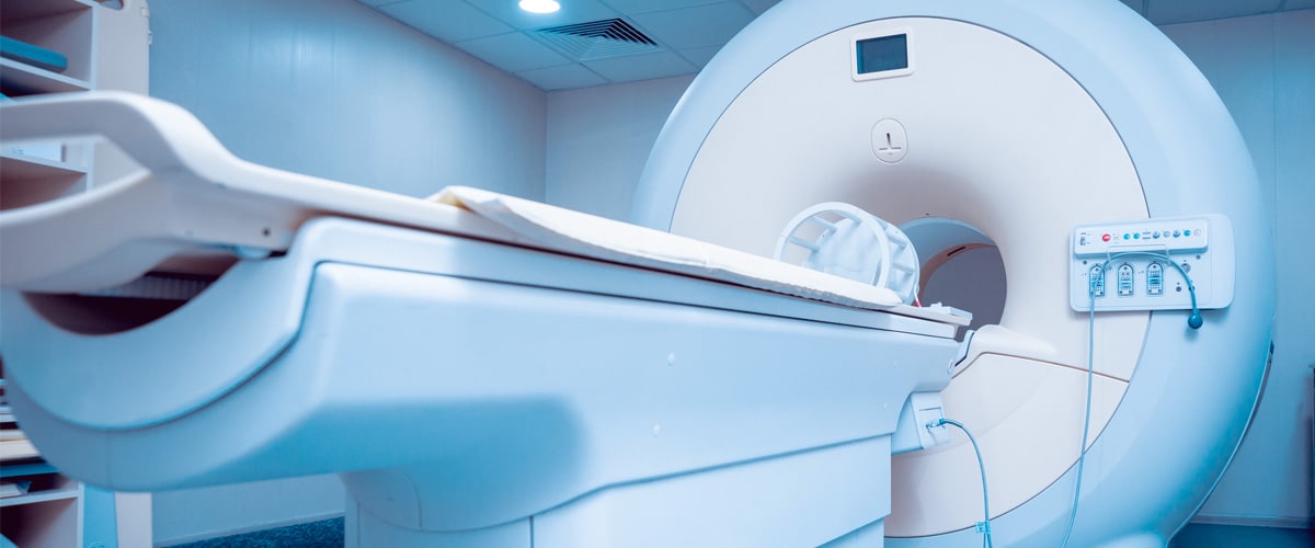
The scan usually lasts between 15 and 30 minutes and are pain-free. Some claustrophobic patients may have initial doubts, but comfort will be found in a detailed explanation of the process and the option of a light sedative.
Our equipment has a high magnetic field, so the quality of the studies is significantly increased and it has the most up-to-date and advanced techniques. It allows for all kinds of studies to be made: nervous system, breast, prostate, heart, vascular, spectroscopy, functional, etc.